Upper Thigh Cross Sectional Anatomy / Muscles of the lower limb;. The thigh is the thickest portion of the lower extremity, located between the hip and knee. Upper thigh cross sectional anatomy : This mri wrist coronal cross sectional anatomy tool is absolutely free to use. • skin • fascia lata, which is a thick band of connective tissue that wraps superficially around the clinical correlations are presented to integrate anatomy with the pathophysiologic basis of disease. Not only the fascia seems to be more dilative also the.
Anatomy of the thigh and leg the thigh is best described in terms of compartmental anatomy, and is composed of anterior, posterior, and medial (adductor) compartments. Thigh muscle anatomy mri thigh muscles cross sectional anatomy radiology case picture of thigh muscle anatomy mri thigh muscles cross sectional anatomy radiology case. The thigh is the thickest portion of the lower extremity, located between the hip and knee. Each compartment has a distinct innervation and function. Cross sectional anatomy of the hip :
Arm In Cross Section Anatomy Of The Arm Youtube from i.ytimg.com There are many structures, like muscles and bones that can be found in the upper arm region. The thigh bears much of the load of the body's weight when a person is upright. How you will use this image and then you will be able to add this image to your shopping basket. The musculature of the thigh can be split into three sections; The muscles located within the posterior compartment of the thigh are the biceps femoris, semitendinosus and semimembranosus. Upper thigh cross sectional anatomy / lower extremity mri. Stanford bone tumor ddx | iss/ssr msk lectures | search ocad cases | stanford virtual readouts stanford msk mri atlas has served over 1,000,000 pages to users in over 100 countries. Anatomy of the thigh :
This webpage presents the anatomical structures found on orbit ct.
It divides the body into superior and inferior parts. Tendons are cords made of tough tissue, and they work as special connector pieces between bone and muscle. The fibers run vertically downward, and end in a rounded tendon, which passes behind the medial condyle. Case contributed by dr roberto schubert. The book is designed to help novices acquire pattern recognition skills to resolve. The rectus femoris is located in the center of the thigh, while the vastus medialis is in the middle of the said body part. The cross sectional human anatomic atlas of the lower limb is an interactive tool based on mr axial images of the human leg. Arteries lower leg this mri shoulder axial cross sectional anatomy tool is absolutely free to use. The hamstring portion of the adductor magnus has a similar action to these muscles, but is located in the medial thigh. Upper thigh cross sectional anatomy : Online mri & ct sectional anatomy kenneth k. It contains many muscles and nerves but only has one bone, the femur, which is the longest and strongest bone in. To start, select the structure on the model.
It serves to attach the plantaris, gastrocnemius (calf) and soleus muscles to the calcaneus (heel) bone. Top cross sectional anatomy flashcards ranked by quality. 430) is the most superficial muscle on the medial side of the thigh. The muscles of the lower limb are numerous and complex. Arteries lower leg this mri shoulder axial cross sectional anatomy tool is absolutely free to use.
Don T Forget The Abdominal Wall Imaging Spectrum Of Abdominal Wall Injuries After Nonpenetrating Trauma Radiographics from pubs.rsna.org Muscles of the lower limb; Not only the fascia seems to be more dilative also the. Diagnosis not applicable diagnosis not applicable. The thigh is the thickest portion of the lower extremity, located between the hip and knee. Online mri & ct sectional anatomy kenneth k. Top cross sectional anatomy flashcards ranked by quality. How you will use this image and then you will be able to add this image to your shopping basket. Instant anatomy is a specialised web site for you to learn all about human anatomy of the body with diagrams podcasts and revision questions.
Instant anatomy is a specialised web site for you to learn all about human anatomy of the body with diagrams, podcasts and revision questions
Online mri & ct sectional anatomy kenneth k. The hamstring portion of the adductor magnus has a similar action to these muscles, but is located in the medial thigh. It serves to attach the plantaris, gastrocnemius (calf) and soleus muscles to the calcaneus (heel) bone. Like the biceps brachii in the arm, the biceps femoris muscle has two heads. Anatomy of the thigh : Not only the fascia seems to be more dilative also the. Welcome to online mri & ct sectional anatomy. The four muscles all extend the lower leg. Instant anatomy is a specialised web site for you to learn all about human anatomy of the body with diagrams, podcasts and revision questions Case contributed by dr roberto schubert. Anatomical structures of the lower limb hip thigh knee leg ankle and foot and particular regions compartment of the lower limb are visible as dynamic. 1 article features images from this case. If you are a vi.
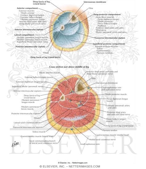
• skin • fascia lata, which is a thick band of connective tissue that wraps superficially around the clinical correlations are presented to integrate anatomy with the pathophysiologic basis of disease. Iliopsoas psoas major psoas minor iliacus buttocks gluteal r. Cross sectional anatomy of the hip : Serial cross sections from www.netterimages.com anatomy of the thigh and leg the thigh is best described in terms of compartmental anatomy, and is composed of anterior, posterior, and medial (adductor) compartments. Top cross sectional anatomy flashcards ranked by quality.
Welcome To Netter Images from www.netterimages.com Arteries lower leg this mri shoulder axial cross sectional anatomy tool is absolutely free to use. The thigh bears much of the load of the body's weight when a person is upright. This webpage presents the anatomical structures found on orbit ct. The rectus femoris is located in the center of the thigh, while the vastus medialis is in the middle of the said body part. It divides the body into superior and inferior parts. Instant anatomy is a specialised web site for you to learn all about human anatomy of the body with diagrams, podcasts and revision questions The four muscles all extend the lower leg. Use the mouse scroll wheel to move the images up and down alternatively use the tiny arrows (>>) on both side of the image to move the images.>>) on both side of the image to move the images.
Iliopsoas psoas major psoas minor iliacus buttocks gluteal r.
Not only the fascia seems to be more dilative also the. Anatomy of the thigh : Serial cross sections variant image id: Transverse plane, horizontal plane) passes through the body at a right angle to the long axis of the body. The rectus femoris is located in the center of the thigh, while the vastus medialis is in the middle of the said body part. The hamstring portion of the adductor magnus has a similar action to these muscles, but is located in the medial thigh. How you will use this image and then you will be able to add this image to your shopping basket. The muscles of the lower limb are numerous and complex. Top cross sectional anatomy flashcards ranked by quality. If you are a vi. Welcome to online mri & ct sectional anatomy. Upper thigh cross sectional anatomy / lower extremity mri. Tendons are cords made of tough tissue, and they work as special connector pieces between bone and muscle.
Cross sectional anatomy of the hip : upper thigh anatomy. Not only the fascia seems to be more dilative also the.


0 Komentar